X


|
STUDENT DIGITAL NEWSLETTER ALAGAPPA INSTITUTIONS |

|
Bob Atkins
https://research.monash.edu/en/persons/bob-atkins
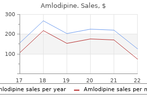
The specific radiation safety issue for each of the common therapies is discussed later in this section arrhythmias in children best 2.5 mg amlodipine. Discharge limits Patients may be discharged only when the remaining activity is less than that prescribed by the local regulatory authority blood pressure medication and weight loss 5 mg amlodipine. This can be estimated using a simple ratio of dose rates at a standard distance referenced to the dose rate immediately following dose administration arrhythmia episode buy amlodipine 10mg, or by measurement of a dose rate alone blood pressure normal limit discount 10 mg amlodipine overnight delivery. This information is often modified to take into account the specific circumstances of each patient blood pressure medication names starting with p safe 5mg amlodipine. Design of therapy areas There are two types of therapy areas inpatient areas and areas where outpatient therapies are administered blood pressure medication list by class discount amlodipine 2.5mg otc. The factors to be considered are: - Types of radiation emitted (photon or particle, or mixed); - the potential for contamination and the degree of the hazard; - the type of waste products generated human excreta, biological waste and general waste and the way they should be handled; - the role of nursing and medical staff in the care of the patient (high or low level of care). Normally, the only difference between therapy areas is in the degree of any shielding required and the issues involved in integrating inpatient areas into a ward, such as access control and toilet facilities. Patient comfort should be catered for by radio, music, television and/or videotape facilities as well as a comfortable (but easily decontaminated) chair. A floor drain is advisable in case of spillage of the therapy radiopharmaceutical. General inpatient therapy guidelines Most inpatient therapies involve 131I, as reflected in the guidelines given below. If radiopharmaceuticals with a low risk of contamination are involved, the guidelines may be suitably modified. No member of staff should enter the therapy room without wearing a radiation monitor. Where digital dosimeters are in use, a record of the dose and the name of the staff member should be kept with the monitors outside the treatment suites. No blood samples, urine or faecal samples should be collected without nuclear medicine approval. As the barrier is crossed on leaving the room, this protective clothing must be removed and placed in the disposal bag provided. Guidelines relating to the patient the following guidelines apply: (a) the patient must be aware of the basic regulations listed below before the administration of a radionuclide. Before therapy, the patient should be given a booklet of common questions and answers. Incontinent patients may need to be catheterized prior to the administration of therapy. Patients should be offered disposable hospital nightwear and towels to prevent their own becoming contaminated. If they wish to wear their own clothes, they must be advised on what should be done with garments on discharge. Ideally, there should be a refrigerator to keep milk fresh, and to store cold drinks if required. This encourages the patient to drink freely and reduces the radiation exposure to nursing staff. Patients should use disposable tissues rather than handkerchiefs, since nasal mucosa tends to have a high radioactive content. Should sheets and/or pillow covers require changing, used linen should be placed in a bag and left within the suite. Under no condition should it be sent to the laundry until checked for contamination. This may involve storage prior to incineration in a licensed incinerator or storage until complete decay of the contamination. Patients should only leave the therapy room for the purpose of a scan or in an emergency, in which case protective clothing. Unless an emergency precludes this, protective clothing should be put on upon leaving the room and removed on re-entry to the suite. Any other belongings that may have become contaminated must be stored for a suitable length of time to allow the radioactivity to decay. The patient should be given a discharge card listing the radionuclide and activity administered, the activity on discharge and any necessary precautions. Contamination With any radionuclide therapy, there is a high potential for contamination. It is, however, strongly advisable to keep a small decontamination kit in or near the therapy area (inpatient or outpatient) for immediate access if required. Radioiodine therapy (a) Pre-therapy It is imperative that a doctor explain to female patients that therapy cannot be given to pregnant patients. If there is any chance that a patient may have become pregnant by the time the therapy administration is to commence, she must report this to a nuclear medicine doctor or technologist. Because of the significantly greater radiation hazards from liquid sources, the comments below assume the use of capsules. In addition to the general advice given above, the following points should be considered when designing the treatment protocol: - Patients should be given written information about the therapy, and in particular instructions for when they return home. The patient may then leave, after any subsequent restrictions are clearly understood. These restrictions may include: - Flushing the toilet twice after urinating, for the first 72 hours after therapy; - Maintaining a safe distance (1 m) from children or pregnant women for a few days. Patients with thyroid cancer will have a very low iodine uptake, and a high proportion (often more than 95%) of the dose will be excreted, generally in the 72 hours following administration. While most excretion occurs in the urine, significant contamination can occur in saliva, with less in sweat and 440 6. Until the dose is fully absorbed from the gut, vomiting can cause a major contamination problem. To deal with these problems, the following measures can be considered: (1) (2) A prophylactic anti-emetic should be given prior to , or immediately after, the dose is administered. The therapist should check with the local regulatory authority for any requirements relating to discharge of radioactive human waste in the sewer. The simplest precaution is to tell the patient to flush the toilet at least twice after urinating. Even then there may still be a requirement (in some countries) to connect the toilet to a storage tank, where the waste may decay for some weeks before discharge to the sewer. If the patient is incontinent, or confused, a bladder catheter should be inserted prior to dose administration. To stimulate excretion, patients should be advised to drink freely, and void frequently. This is a short information sheet to help you understand the restrictions that will be placed on you after undergoing treatment using radioactive iodine. There are several precautions that you and your family must observe both during the time you are in hospital and after you have been discharged. These precautions must be discussed fully with you; they are outlined below to ensure that they are clear. The radioactive treatment cannot be administered unless you understand these restrictions and sign a consent form by which you agree to adhere to them. Since you will become radioactive and will emit radiation after the treatment, you will be required to remain within the radionuclide treatment room until you are advised that it is safe to leave. You will excrete a considerable amount of radioactive iodine in urine, faeces, sweat, saliva and nasal mucous. It is very important that these substances are not allowed to contaminate other people, or areas outside the room. You will be provided with an electric kettle, coffee powder and tea bags so that you may make your own drinks. Money: It is not advisable to bring more money than you think you will require into the ward. If you would like the nursing staff to buy you daily newspapers, please give them some cash prior to the initiation of your treatment. If you are likely to have much excess money, it is wise to ask the nursing staff to lock it away until it is time for you to go home. Clothes: Any clothes that you wear may become contaminated with radioactive iodine. Ideally, clothes worn while in the ward should be suitable for laundering in a washing machine; they should be taken home in a polythene bag and washed in a machine. Other personnel belongings: It is advisable not to bring too many personal belongings into hospital, since anything you handle could become contaminated. If you are on any medications, including nose drops, eye, ear, throat or cough drops or tablets, you must inform your doctor since they could prevent the radioactive treatment from acting efficiently. It is important for you to drink as much fluid as possible, as this helps to keep the radiation dose to the bladder to a minimum, thus preventing a possible cystitis. It is also important not to be constipated, since this will lead to the stomach and bowel becoming unnecessarily irradiated. As a result the residual whole body activity will remain high for a longer period, possibly delaying your discharge. You should use disposable tissues rather than handkerchiefs, if possible, since nasal mucous tends to have a high radioactive content. Bed linen: Should this require changing, the used linen should be placed in a bag and left within the suite. Any radioactive contamination can normally be washed out in one washing machine cycle, but this must be done under the control of a physicist. It is permitted for you to have visitors, provided that they comply with the regulations and that neither children nor pregnant women visit you at any time. Under no circumstances should visitors eat, drink, smoke or use the toilet facilities in the treatment room. There are different levels of remaining activity at which the hospital is allowed to discharge you depending on your home circumstances and on your means of transport home. After two or three days (possibly longer, depending on the estimate of residual activity), the activity remaining within you will be measured and you will then be advised as to how many more days it may be necessary for you to stay. Once you have been discharged, we may have to request you to observe certain restrictions such as not going to the cinema or mixing with children. In the event of an emergency, the nuclear medicine department should be contacted immediately (be sure to include contact information). Pure beta emitters Beta emitters such as 186Re, 169Er, 166Ho, 90Y, 89Sr and 32P generally require consideration only at the time of administration, with little if any hazard afterwards, although higher energy beta emissions can cause measurable bremsstrahlung X ray radiation exterior to the patient. Administration must only be performed in a room designed for unsealed radionuclide use; in particular, all surfaces should be free of gaps and easily washable. Following administration the injection site must be checked for spilt or leaked radionuclides by swabbing and checking the swab with a beta detector. In the case of 89Sr, most of the unbound strontium will be excreted in the urine, normally within 48 hours of administration. Precautions must be taken if staff or relatives come into contact with urine, for example if the patient is incontinent or catheterized, in which case gloves must be worn and properly disposed of. Patients should again be advised to flush the toilet twice after voiding for the first 48 hours. The patient must be given written instructions covering the need to wash hands following toilet use, cleaning up any spilt urine, flushing toilets and the washing of any clothing that may be urine contaminated. The instructions should also include contact names and phone numbers in case of an emergency. Emitters of both beta and gamma radiations Samarium-153 and 188Re produce some gamma emission but this is not normally a significant safety problem. Staff and patient should be advised to remain at a distance of 1 m until discharge. Precautions following death of a therapy patient Procedures should be put in place for the safe disposal of the bodies of patients who have received therapeutic doses. This may include labelling, contamination avoidance and notification of the staff who may have to handle the body. These procedures will obviously depend on the radiopharmaceutical involved, the dose and the time since administration. If a post-mortem or embalming is to be performed, then advice from the radiation safety officer must be sought, as there could be a significant contamination and radiation hazard, especially with 131I. Ward staff in particular should be provided with instructions for dealing with the death of a radionuclide therapy patient. Procedures the nuclear medicine department must be notified as soon as possible after a death. The body should be removed from the ward to the mortuary as soon as possible after death, without attempting to remove any of the radioactive material, and placed in, if possible, the centre section of the body storage refrigerator. This is to minimize any radiation exposure to staff who may be working in the mortuary. Mortuary procedures Exposure of individuals to radiation emitted by radioactive materials retained in or on a corpse can be reduced by: (a) (b) (c) Working quickly to reduce the time of exposure; Working, where necessary, behind adequate shielding; Consulting with nuclear medicine staff for advice on radiation safety and removal of highly contaminated tissues such as the thyroid. If, however, a corpse contains radioactivity in excess of the levels given above, the pathologist should be informed of the radiation levels likely to be 446 6. Occasionally corpses are assigned to medical schools for dissection or are to be transported overseas. Any hazards to persons involved in these operations or the need for compliance with international transport regulations depend on several factors relating to the nature of the radioactive sources. In most instances the issue is resolved by keeping the corpse in appropriate cold storage until twenty half-lives of radioactive decay have passed. If it is known that the radioactive material used for treatment will be selectively absorbed in a particular organ, for example 131I in the thyroid, the organ should be excised before the examination proceeds and removed from the work area. If it is known that radioactive material used for treatment will be distributed in particular body fluids, these should be drained off, using suitable equipment, before the examination proceeds. The equipment should later be decontaminated by thorough rinsing in a detergent solution followed by washing in running water. Transport of a corpse containing radioactive materials should be considered in accordance with the requirements of local legislation covering the transport of radioactive materials. Cardiac or respiratory arrest, or transfer of a therapy patient for medical reasons Resuscitation of patients containing radioactive material for radiotherapy or therapeutic nuclear medicine purposes poses special problems.
Although the number of animal outbreaks between 1970 and 1979 had declined to almost a quarter of that of the previous decade (19601969) arteria3d unity buy amlodipine 5mg with amex, there were 300 outbreaks a year blood pressure 3 year old cheap amlodipine 5mg with amex, mostly involving sheep blood pressure pictures buy generic amlodipine 5 mg on-line. During this period hypertension xanax order amlodipine 10mg with visa, there were 8 arrhythmia nursing diagnosis generic amlodipine 5mg free shipping,475 sheep and 1 blood pressure chart free printable discount amlodipine 10 mg with mastercard,675 bovine losses in 3,669 separate outbreaks. During the same period, 482 human anthrax cases occurred in the country and all patients were from rural areas [6]. The highest incidences were observed in the prefectures of Aetoloakarnania, Evros, Ioannina, Larissa, Rodopy and Thessaloniki [6]. Since then, strict control measures have eliminated the disease and only sporadic cases in animals and humans have been reported. Thessaly is a rural region located in Central Greece and includes four prefectures (Karditsa, Larissa, Magnesia, Trikala). The large majority of them (more than 1 million goats and sheep) are farmed in Larissa prefecture. According to the records of the local Veterinary Authority of Larissa, three outbreaks of anthrax have been reported in Larissa in the past 35 years (in 1978, in 2000, and in 2006) (unpublished 11 Epidemiological investigation the stockbreeder was contaminated after having handled the slaughtered sheep due to direct contact with the infected animal. He had flayed the animal together with his wife and then fed two dogs with the contaminated meat. However, she did not present any signs or symptoms of infection and is now under post-exposure prophylactic treatment. The residents of the village (Tsabournia) have been informed about this case in order to recognise early clinical symptoms of anthrax and they were advised to seek medical treatment immediately if anthrax was suspected. The local health centre and general practitioners are aware of this need for careful monitoring. Special directions have been given to the stockbreeders of Tsabournia regarding the use of protective equipment. The local Veterinary Authority has taken measures for the correct disposal of animal carcasses, including disinfection of contaminated material and All of them occurred in herds kept in two villages (Livadi and Tsabournia) situated at a distance of 35 km from each other in the area of Elassona, Larissa prefecture. Approximately 90 animals were affected in total, and the outbreaks were contained after correct disposal of animal carcasses and vaccination of exposed animals. According to the epidemiological data of the Veterinary Authority, no case of anthrax in animals or humans has ever been declared in the other three prefectures of Thessaly. In 1978, anthrax infection had been confirmed in animals of three different herds in Tsabournia. Vaccination and appropriate control measures have been taken; since then until the incident described here no other anthrax case in animals or in humans has been reported. ArticleId=19464 Holzmann T, Frangoulidis D, Simon M, Noll P, Schmoldt S, Hanczaruk M, et al. Anthrax infection among heroin users in Scotland during 2009-2010: a case-control study by linkage to a national drug treatment database. Karpouzis A, Panopoulou M, Bazzano G, Grapsa A, Maltezos E, Ktenidou-Kartali S, et al. Conclusions From a public health point of view, anthrax is important for Europe as well as for other regions. Here, post-exposure prophylaxis was nevertheless recommended after hospital discharge because the precise conditions of direct contact which took place during flaying were not clearly known [17]. Early recognition of this suspected human case and reporting to the local authorities without delay have led to the prevention of further spread of the disease both in humans and animals. French Ministry of Agriculture, Agro-food Industry and Forest, General Directorate for Food, Paris, France 3. Regional office of the French Institute for Public Health Surveillance, Lyon, France 6. Associate national reference laboratory, Microbiology department, University hospital Caremeau, Nimes, France 8. ArticleId=20227 Article submitted on 13 July 2012 / published on 26 July 2012 A case of human brucellosis was diagnosed in France in January 2012. The investigation demonstrated that the case had been contaminated by raw milk cheese from a neighbouring dairy farm. As France has been officially free of bovine brucellosis since 2005, veterinary investigations are being conducted to determine the origin of the infection and avoid its spread among other herds. In January 2012, a human case of brucellosis was diagnosed by blood culture in a district of the French Alps. The patient had presented in late November 2011 with non-specific symptoms that had been ongoing since that date. Usual at-risk exposures were investigated: recent or ancient travel in an endemic/enzootic country, consumption of raw milk or raw milk products imported from an enzootic country, professional or accidental exposure to Brucella strains in a laboratory, direct contact with animals, etc. As the patient had not had such an exposure at any point before, the case was considered to be an autochthonous case of acute brucellosis of undetermined origin. In April 2012, brucellosis was confirmed in a dairy cow in a herd of the same district of the French Alps. The seropositive cow had aborted in late January, and a strain of Brucella melitensis biovar 3 was isolated from the milk sampled from the animal. The animal belonged to a herd 21 dairy cows, and no other animal in the herd presented with symptoms suggesting brucellosis or showed any serological reaction. Approximately 20 kg of Reblochon cheese (soft raw milk cheese) are usually produced daily on the affected farm. Brucellosis surveillance in France France has been officially free of brucellosis in cattle since 2005, and the last outbreak of brucellosis in sheep and goats was reported in 2003. Moreover, abortion in ruminants is mandatorily notifiable and the investigation of abortion includes examination for brucellosis. The confirmation is carried out using a combination of in-house tests including Rose Bengale test, immunoassay, complement fixation test, and specific detection of antibodies against Yersinia enterocolitica. All animals were tested serologically (Rose Bengale test, complement fixation test and indirect enzyme linked immunosorbent assay) before slaughter in April [5]. All animals were seronegative with the exception of the index animal which showed a very strong reaction in all three tests. Following the confirmation of brucellosis in the cow, a trace-back investigation was implemented by the veterinary services to determine the origin of the contamination of the herd. The animals of the infected herd had not taken part in a transhumance nor did they graze with other herds on the same pastures. Other neighbouring farms as well as farms that had traded animals with the infected farm in the year before the outbreak were investigated. A trace-forward investigation was also carried out to determine the places of distribution of cheese produced at the affected farm since the abortion of the cow. Reblochon cheese is a raw milk soft cheese, requiring a maturation period of three weeks to one month. The cheese from the affected farm had been commercialised after the abortion in seven districts. Cheese was sold directly at the farm, and as whole pieces or in parts in supermarkets. Cheese produced by the affected farm had not been exported to other countries but might have been bought by foreign tourists during their winter holidays in several ski resorts in the area. Consumers of these products were advised to seek medical attention should they present symptoms consistent with brucellosis. The movements of animals from other herds that had epidemiological links with the infected herd (those that were geographically close to the infected herd, or had been bought from the infected herd) have been restricted until the end of the investigation. Furthermore, raw cheese products from farms with epidemiological links to the infected farm were put on sale only after negative bacteriological tests results had been obtained. Reinforcement of human surveillance Notification of human brucellosis is mandatory in France. All notified human cases in France have to be confirmed by the national reference laboratory. Among them, 183 (84%) were patients infected through the consumption of raw milk products or direct contact with animals in (or from) countries with enzootic brucellosis, 14 (6%) were laboratory workers infected through the handling of Brucella strains, 17 (8%) were relapses in people with past infection, while the origin of contamination could not be determined for five patients (2%) [9]. Because the investigation of the origin of the human case diagnosed in January 2012 had been inconclusive, it was decided to reinforce the surveillance immediately. Since January 2012, all notified suspected cases have been interviewed with a trawling questionnaire before the diagnosis was confirmed. Since April 2012, any epidemiological link with the infected herd has been systematically investigated. Human investigations After the identification of the first bovine case, the human case was interviewed again to investigate any direct or indirect epidemiological link with the infected herd. During the second interview, it became clear that the patient and their family had visited the infected farm in autumn 2011, although it was not possible to determine the exact date. During this visit, the family had bought Tome Blanche cheese, a fresh cheese obtained during the first step of Reblochon production. The four family members had shared the Tome Blanche on the same day, but the index case was the only one who later presented with symptoms. The other three family members were serologically investigated in May 2012 and only one presented with a positive high titre in agglutination (1,600). Microbiological investigations the strain isolated from the human case and from the two cows both belonged to Brucella melitensis biovar 3. Control measures All cheese pieces produced by the affected farm and still within the shelf life were withdrawn from retailers. In addition, a recall of already sold products was carried out via a national press release by the cheese producer and by posters in the sale points. Medical 14 At this time, several hypotheses can be proposed to explain the re-emergence of brucellosis in cattle in France. Knowing that the affected herd had not received any imported animals, it needs to be investigated whether animals had been introduced in one of the herds that sold animals to the affected farm or whether the affected herd had been in contact with animals of neighbouring farms. However, no infected chamois has been identified in the last 10 years, despite several serological surveys (Garin-Bastuji, personal communication, July 2012). The veterinary investigations are still ongoing to determine the origin of the contamination of the herd, to investigate the possible spread of the infection to other herds and to take control measures to avoid the infection of new herds and consequently the occurrence of additional human cases. However, the absence of infected animals in the herds that are epidemiologically linked with the infected herd, and the absence of other autochthonous human cases argue in favour of a single outbreak and a limited episode. The index animal on the farm was born from a dam that itself was born in 1999 before the last outbreak in the area and died in 2006. The lifetime of the mother of the index infected animal is therefore consistent with the hypothesis of a congenital case of bovine brucellosis [11]. In addition to the investigations already carried out, all herds coming back from transhumance in the concerned district will be serologically screened during the fall. Serological tests lack specificity but they have a good sensitivity and are of good value to detect recent or active infections. The index animal had an active infection demonstrated by Brucella excretion in milk. This animal displayed a high level of antibodies in relation with the active although possibly chronic infection. Strengthened surveillance of human and animal brucellosis will be maintained until the end of the investigations. The surveillance of human brucellosis in non-endemic countries is complicated by the lack of specificity of serological tests [12-16]. In our experience, all available tests still may cross-react with other bacteria (mainly Y. In countries with low prevalence and incidence of the disease, this low specificity contributes to the low positive predictive value of serology. A positive diagnosis has important consequences for the patients (long antimicrobial therapy with possible adverse effects and ecological consequences on intestinal bacteria), and for the dairy animals (culling of the entire herd in our country). It is therefore important to obtain as much evidence as possible to confirm a serological diagnosis. Isolation of Brucella melitensis biovar 3 from а chamois (Rupicapra rupicapra) in the Southern French Alps. False positive seroreactivity to brucellosis in tuberculosis patients: a prevalence study. Evaluation of in-house and commercial immunoassays for the sero-diagnosis of brucellosis in a non-endemic low prevalence population. Specificity dependence between serological tests for diagnosing bovine brucellosis in Brucellafree farms showing false positive serological reactions due to Yersinia enterocolitica O:9. Efficacy of several serological tests and antigens for diagnosis of bovine brucellosis in the presence of falsepositive serological results due to Yersinia enterocolitica O:9. Investigations and actions taken during 2011 due to the first finding of Echinococcus multilocularis in Sweden. ArticleId=20215 Article submitted on 6 February 2012 / published on 12 July 2012 Echinococcus multilocularis is a parasite that can cause alveolar echinococcosis disease. In this group, all relevant information was shared, strategies for information dissemination and any actions to be taken due to the finding of E. The present paper describes the actions taken during 2011 and the results thereof, including surveillance in animals, risk assessment for humans to become infected and recommendations given to the public. Further discussion about whether the parasite was introduced, and if so, how, as well as possible future development of the infection in animals and humans in Sweden and future actions are included. It is considered to be the most serious parasitic disease in humans in Europe [1]. The parasite develops with a tumour-like growth almost exclusively in the liver and the disease is characterised by a long incubation period, between five and 15 years, followed by a subsequent chronic course [2]. Although a serious disease, in Europe, the reported prevalence in humans is low, up to 1.
Positive inotropes increase myocardial contractility arrhythmia beta blocker discount amlodipine 2.5mg with visa, while negative inotropes decrease myocardial contractility blood pressure medication used for adhd generic 2.5mg amlodipine overnight delivery. Surgical Terms T Surgery is the branch of science that treats diseases blood pressure under stress buy 10 mg amlodipine amex, injuries arrhythmia 4279 buy 5 mg amlodipine otc, and deformities by manual or operative methods blood pressure 70 over 50 purchase 10mg amlodipine with visa. Some surgical terms include the following: appositional (ahp-sih-shuhn-ahl) = placed side to side blood pressure wrist monitor purchase amlodipine 10 mg online. An allograft (ah-l-grahft) is a graft from another individual of the same species. An isograft (-s-grahft) is a graft from genetically identical animals, such as twins or inbred strains. Drugs, Diseases, and Dissection 357 postop (pst-ohp) = common term for after surgery; postoperatively. Resect is used in reference to holding tissue or an organ out of the surgical field. Transfixion sutures are used for large, isolated vessels and organs to prevent slippage of the ligature. Surgical Equipment autoclave (aw-t-klv) = apparatus for sterilizing by steam under pressure (Figure 1712). Drugs, Diseases, and Dissection Tip Jaw Boxlock Boxlock pin 359 Shank Ratchet Ring handle Ratchet Figure 1715 Parts of surgical instruments. Tibia Intramedullary pin (a) Cerclage wire Figure 1716 Radiograph showing the repair of a fractured tibia. The fluid does not drain through the center of the tubing, but is allowed to leak from the skin openings. Hand chuck Ronquer Gelpi retractor Kern bone holding clamp Periosteal elevator (a) Emasculator Emasculatome Scalpel handle Halsted mosquito hemostatic forcep Adson Brown thumb tissue forcep Surgical Approaches In surgery, the specific procedure by which an organ or a part is exposed is called the approach (ah-prch). Following are examples of different surgical approaches (Figure 1720): flank incision (flahnk ihn-sihz-shuhn) = surgical cut perpendicular to the long axis of the body, caudal to the last rib. The first definition is more correct; however, the term biopsy is commonly used both ways. Types of biopsies include the following: excisional biopsy (ehcks-sih-shuhn-ahl b-ohp-s) = removing entire mass, tissue, or organ to examine. Drugs, Diseases, and Dissection 361 A necropsy (n-krohp-s) is a postmortem examination that consists of a thorough examination of a dead animal to determine the cause and manner of death and to evaluate any disease or injury that may be present. Needles and Sutures Suture material and needles are used by surgeons to close wounds or to tie things (Figure 1721). Terms used in reference to suture material and needles include the following: blunt (bluhnt) = dull, not sharp; used to describe needles or instrument ends. Ventral midline incision Paramedian incision Skin stapler Flank incision Paracostal incision Taper edge needle Cutting edge needle Hemo clips Figure 1720 Incision types. Swaged needle needle biopsy (n-dahl b-ohp-s) = insertion of a sharp instrument (needle) into a tissue for extraction of tissue to be examined. Movement of water across a selectively permeable membrane along its concentration gradient is a. A device by which a channel may be established for the exit of fluids from a wound is a a. A solution that is less concentrated than what it is being compared with is known as a. The graded locking portion of an instrument located near the finger rings is the a. Originally, dogs and cats were domesticated for work such as herding and Or controlling rodents. Although dogs and cats still may be used for work, they con are more commonly kept as pets. Many of the anatomy and physiology concepts and medical terms related M to dogs and cats have been covered in previous chapters. The lists in this chapter apply more specifically to the care and treatment of dogs and cats. Elizabethan (-lihz-ah-bth-ahn) collar = device placed around the neck and head of dogs or cats to prevent them from traumatizing an area; commonly called an E-collar (Figure 184). Ruptured anal sac abscess Gingivitis Figure 181 Line drawing of anal sac location. Breed-Related Terms angora (ahn-gr-ah) = type of long fur on cats (and other species). Crotalus atrox toxoid (kr-tah-luhs ah-trohcks tohcks-oyd) = inactivated toxin from the Western diamondback rattlesnake used in dogs to reduce morbidity and mortality due to envenomation by this snake. Giardia lamblia (g-ahr-d-ah lahmb-l-ah) = protozoan that may cause asymptomatic disease or cause diarrhea in dogs and cats. Lyme (lm) disease = bacterial disease caused by the bacterium Borrelia burgdorferi transported by a tick vector; associated with fever, anorexia, joint disorders, and occasionally neurologic signs; also called Lyme borreliosis. The pouches that store an oily, foul-smelling fluid in dogs and cats are called a. What is the device placed around the neck and head of dogs and cats to prevent them from traumatizing an area? A 3-yr-old F/S black Labrador retriever was presented to the clinic for removal of a round bone from the mandible. Hx: the dog had been chewing on the bone during the day and had gotten the bone stuck on its mandible. Gigli wire was threaded through the hole in the center of the bone, and the bone was cut in two places to allow its removal. While the bone was being sawed, tissue trauma occurred to the skin of the mandible. Abdominal palpation yielded normal kidneys, normal intestinal loops, a tense and painful caudal abdomen, and a turgid urinary bladder. When the bleeding was under control, the dog was anesthetized so that the blood vessels could be ligated and the wound sutured. When the abdominal incision was being closed, the veterinarian noted pooling of blood in the abdomen. A large amount of blood was coming from the abdominal incision, and the veterinarian had the technician reassess the animal. The owner was called to see whether the dog had been sick recently, and the owner stated that the dog was seen eating rat bait about 3 days earlier. Additional blood was collected in a heparin tube for assessment of clotting times, and the dog was given vitamin K1. The dog made a slow recovery and was hospitalized an additional night for observation. The dog was discharged with antibiotics, and an appointment was made to reassess the anal sacs in 7 days. Horses, donkeys, mules, and ponies have been used as companion rec animals as well. The lists in this chapter apply more specifically to the care and treatment of equine species. Western (wehs-trn) = method of riding in which the stirrup length is long, the rider rides in an upright posture, and the rider has a one-handed hold on the reins. Monkey mouth (lower incisors protrude beyond upper incisors) Parrot mouth (upper incisors protrude beyond lower incisors) Figure 192 Types of bad mouth (malocclusions). Tip of coffin bone In chronic founder, the coffin bone is detached and rotated toward the sole. Star Stripe Snip Star and stripe Blaze Star, stripe, and snip Bald Chin spot Star and snip Spot Race Half-heel Heel Coronet Half-pastern Pastern Sock or ankle Halfstocking Stocking Figure 197 Natural face and leg markings. Scotch hobble (skohtch hohb-uhl) = type of restraint in which all four feet are tied in an X pattern to keep them from moving or kicking. Management Terms as-fed basis (ahs-fehd b-sihs) = amount of nutrients in a diet expressed in the form in which it is fed. In longeing, a horse on a long strap or line travels in a large circle around the handler. Longeing helps train young horses, exercises horses, and improves balance and development of stride. English saddle Side view Cantle Seat Pommel Skirt Panel Stirrup bar Stirrup iron Tread of stirrup iron Skirt Backhousing or back jockey Cantle Horn Fork Seat Pommel Western saddle 387 Wool lining Rope strap Lace string Skirt Dee ring Front jockey and seat jockey, one piece Flap Stirrup leather Lace strings Dee ring Bottom view Panel Sweat flap Flap Leather flank girth billet Stirrup leather keeper Stirrup leather Front tie strap or cinch strap Fender or sudadero Stirrup Billets Figure 1913 Parts of the English saddle and Western saddle. Types of Horses draft (drahft) horse = large breed of working horse (usually over 17 hands) (Figure 1914). Equine Vaccinations equine encephalomyelitis (ehn-sehf-ah-l-m-ih-ltihs) = mosquito-transmitted infectious alphaviral disease of horses that is associated with motor irritation, paralysis, and altered consciousness; there are three types: Eastern, Western, and Venezuelan; also known as sleeping sickness. Potomac (p-t-mihck) horse fever = rickettsial bacterial disease of horses that is associated with fever, anorexia, incoordination, diarrhea, and edema of the extremities; also called equine ehrlichiosis (ehrlhck-sihs). Poll evil Knocked-down hip or capped elbow Fistulous withers 389 Stifled Thoroughpin Sweeney Capped hock Bone spavin Bog spavin Splint Ringbone Bowed tendons Quittor Grease heel (scratches) Toe crack Quarter crack Laminitis (founder) Osselets Splint High ringbone Low ringbone Figure 1916 Terms for unsoundness in horses. West Nile virus = mosquito-borne viral encephalitis that entered the United States (probably from the Middle East) in 1999. A person who cares for equine feet, including trimming and shoeing, is called a(n) a. In horses, a rapid two-beat gait in which the front and hind feet on the same side start and stop at the same time is called the a. What is the term for the large, air-filled outpouching of the eustachian tube in equine? The vice in horses in which they grasp an object between the teeth, apply pressure, and swallow air is a. The canine tooth in a horse that is usually found only in males also is called the a. A 6-yr-old quarter horse mare was presented for signs of pawing at the abdomen, flank-watching, anorexia, and lack of stool production. The veterinarian did a rectal palpation of the horse and noted gas-filled segments of bowel that felt proximal (oral) to an impaction. Upon palpation of the hoof, the area near the coronary band was warm and a pulse could be palpated. The first three-the rumen, reticulum, and omasum-are com actually outpouchings of the esophagus. Dairy cattle are bred for their milk-producing qualities, whereas beef cattle are bred for meat. Some breeds mil are considered dual purpose, which means they have both dairy and beef traits. The lists in this chapter apply c more specifically to the care and treatment of cattle. F1 generation (F-1 jehn-r-shuhn) = first offspring from purebred parents of different breeds or varieties; F1 stands for first filial. Milk-Related Terms alveoli (ahl-v-l) = milk-secreting sacs of mammary gland; also used to describe gas exchange sac of respiratory system. Make Room for the Ruminants 399 Switch Switch Figure 207 the distal part of a bovine tail is the switch. Alveoli Interior of alvelous showing milk producing cells Milk ducts Gland cistern Teat cistern Milk cavity Streak canal Circular muscle (sphincter) Tubule Fat droplets in lumen Blood supply Figure 209 Parts of the udder. As in cattle, some breeds are better known for their wool production and others are better known for their meat quality. Many of the anatomy and physiology concepts and medical terms related to sheep have been covered in previous chapters. The following list of words is used more specifically for sheep and sheep production. Make Room for the Ruminants 403 Figure 2018 Scouring removes material from the wool. Many of the anatomy and physiology concepts and medical terms related to goats have been covered in previous chapters. Additional terms pertaining to goats are listed under the cattle section because they apply to both animals. Camelids are considered pseudoruminants because they have three stomach compartments (reticulum, omasum, and abomasum) instead of four. There are three genera of camelids; the true camels of Asia (genus Camelus); the wild guanaco and the domesticated alpaca and llama of South America (genus Lama); and the vicuсa of South America (genus Vicugna). Llamas are the larger species with less desirable wool; alpacas are smaller than llamas and have high-quality wool; vicunas are undomesticated and are the smallest and rarest with fine, high-quality wool; and guanacos are slightly smaller than llamas and are undomesticated. Llamas are becoming popular pets and are used as pack animals and for fiber production (Figure 2020). Many of the anatomy and physiology concepts and medical terms related to llamas have been covered in previous chapters. Hemophilus somnus (h-moh-fihl-uhs sohm-nuhs) = bacterial infection that causes reproductive, urinary, respiratory, and septicemic disease in cattle. Make Room for the Ruminants 405 leptospirosis (lehp-t-sp-r-sihs) = bacterial disease caused by various serotypes of Leptospira; signs include renal failure, jaundice, fever, and abortion. The inedible visceral organs and unusable tissues removed from the carcass of a slaughtered animal are the a. What is the name of the passageway that takes milk from the teat cistern to the outside of the udder? What is the term for the restraint device that secures cattle around the neck to allow accessibility for milking, feeding, and examining? The behavior in which a camelid propels saliva at an animal or a person who is perceived to be a threat is called a.
When the bladder becomes full hypertension table order amlodipine 2.5 mg mastercard, urethral sphincters are opened then urine passes through urethra to be voided out pulse pressure compliance amlodipine 5mg. Clinical Features - Acute urinary retention - Symptoms of prostatism (frequency blood pressure entry chart cheap amlodipine 5mg with amex, urgency blood pressure chart dogs cheap 5 mg amlodipine with amex, dribbling blood pressure medication regimen buy amlodipine 10mg mastercard, dysuria exo heart attack buy discount amlodipine 2.5mg, etc) - Chronic retention insufficiency. Classification Benign Renal Tumors - Not very common - Includes: Adenoma of renal parenchyma - Papilloma of renal parenchyma - Lipoma, etc - Hematuria and dull pain in the flank are common complaints. Malignant renal tumors - Include: - wilms tumor: - it occurs in children - Renal cell carcinoma: - it occurs in adults - Clinical features - Pain and hematuria are the earliest symptoms. Post procedural urethral stricture:It occurs following urologic procedural like catheterizations. Minor trauma to the urethral mucosa may heal by forming strictures years after the procedure. Urine stasis and back-flow of urine: - Results in hydronephrosis and Renal- failure Symptoms of renal-failure (uremia). Pathophysiologic Effects of Obstructive Uropathy Obstruction to urinary path way (flow) Stasis of urine and back flow Bacterial over growth (pylonephiritis) 200 Pathophysiology Hydronephrosis distension of the renal pelvis. Small increase in one component can be compensated in decrease in volume of one or both of other components of the cranial cavity. Normal fluctuation in intracranial pressure occurs with respiratory movement and activities of daily living such as straining, coughing and sneezing. Example: o An increase in tissue volume results from brain tumors, brain edema, or bleeding into brain tissue. When the pressure in the cranial cavity approaches or exceeds the mean systemic arterial pressure, tissue 205 Pathophysiology perfusion becomes inadequate, cellular hypoxia results and if maintained, neuronal death may occur. The level of consciousness may deteriorate from alertness through confusion, lethargy, obtundation, stupor, and coma. Neurons in vasomotor centre respond directly to ischemia by producing a marked increase in mean arterial blood pressure. Some times up to 270mmHg is accompanied by widening of pulse pressure and a reflex slowing of the heart rate. Types of Brain Edema:208 Pathophysiology There are two types of brain edema:a) Vasogenic Brain Edema:- Increased extra cellular fluid that surrounds brain cells. Infratentorial Herniation Supratentorial Herniation There are three subtypes based on sites. Infratentorial herniation:Results from increased pressure in the brain compartment and often progress rapidly. Cause death because it is likely to involve the lower brain stem centre that control vital function. Brain Injuries Injury to the brain parenchyma:- There are two types of brain injury:a) Primary Head injury b) Secondary Head injury a) Primary Head Injury: 213 Pathophysiology Primary head injury the impact. The causes are: Intracranial hematoma Brain Edema, and Infections Intracranial Hematoma Is bleeding in the cranial cavity. There are four common types based on the site of bleeding:· · · Epidural hematoma:- bleeding outside dura Subdural hematoma:- bleeding below dura Subarachinoidal hematoma:bleeding below the arachnoids matter. Infections:- 215 Pathophysiology Intra cranial infection one of a common and fatal condition. There are two types of intracranial infections:a) Meningitis: - infection of Meninges b) Encephalitis: - infection of brain parenchyma. Classifications: · · · Acute Meningitis Causes · Acute pyogenic Meningitis: bacterial -Streptococcus pneumonia. Most common symptoms and signs: · Fever, chills: - due to release of inflammatory mediators and endotoxins. Brudzinski sign: when forcible flexion of the neck results in flexion of hip and knee. B the path physiologic mechanisms of these signs are due to stretching of inflammed meninges, which results in pain upon performing the maneuvers. A genetic disorder can involve a single gene trait or it can involve a polygenic trait. The effects of an abnormal genetic trait may present at birth or may not become apparent until later in life. The auto somes on each chromosome are arranged in strict order, with each gene occupying a specific location or locus, and in pairs, with one maternal and one paternal member. If both members of a gene pair are identical then the person is homozygous for the locus; if they members are different, then the person is heterozygous. Eg If the trait is only expressed in the hetrozygote, it is said to be dominant and if it is only expressed in the homozygote, it is recessive. In autosomal dominant disorders, a single mutant allele from an affected parent is transmitted to an offspring regardless of sex. The unaffected relatives of the parent or unaffected siblings of the offspring do not transmit the disorder. The affected individual has a 50% chance of transmitting the disorder to each offspring. Autosomal dominant disorders are characterized by reduced penetrance and variable expressivity, and age of onset that is later in life (eg. Although there is a 50% 222 Pathophysiology chance of inheriting a dominant genetic disorder, there can be wide variation in gene expression. Autosomal recessive disorders are manifested when both members of the gene pair are mutant alleles. In this case, both parents may be unaffected but are carriers of the defective gene. The occurrence risk in each pregnancy is one in four for an affected child, two in four for a carrier child, and one in four for a normal (non carrier, unaffected) homozygous child. With autosomal recessive disorders, the expression of the gene tends to be more uniform than with autosomal dominant disorders; the age of onset is frequently early in life, and in many cases enzyme proteins are affected by the mutation. The common pattern of inheritance is one in which an unaffected mother carries one normal and one mutant allele on the x chromosome. When the affected male procreates, he will transmit the defect to all of his daughters, who will then become carriers of the mutant gene. Since the genes of the Y chromosome are unaffected, the affected male will not transmit the defect to any of his sons and they will not be carriers or transmit the disorder to their children. These biochemical defects involve the formation of abnormal structural proteins, abnormal biochemical mediators or enzymes, or abnormal membrane bound transport system or receptor proteins. Structural protein defects are usually manifested as autosomal dominant disorders. Characteristics of the skeletal defects are a long thin body hyperextensive joints, arachnodactyly (spider fingers), and scoliosis. Defects of the eye include the upward 224 Pathophysiology displacement of the lens and the potential for retinal detachment. Involvment of connective tissue in the cardiovascular system may lead to mitral valve disease and a tendency for development of a dissecting aortic aneurysm. In albinism, the basic biochemical defect is the absence or non- functioning of the enzyme tyrosinase. This enzyme is necessary for the production of melanin, the pigment that gives skin its color. In this disorder, there is a deficiency of phenylalanine hydroxylase, the 225 Pathophysiology enzyme needed for conversion of phenylalanine to tyrosine, and as a result of this deficiency, toxic levels of phenylalanine accumulate in the blood. Infants with the disorder are treated with a special diet that restricts phenylalanine intake. Infants with Taysachs appear normal at birth but begin to manifest neurologic signs at about 6 months of age. These neuralgic manifestations eventually lead to muscle flaccidity, dementia, and finally death at about 2 to 3 years of age. Alterations In Chromosome Duplication Mosaicism is the presence in one individual of two or more cell lines characterized by distinctive karyotypes. Alterations in Chromosome Number A change in chromosome number is called aneuploidy. Among the causes of aneuploidy are failure of separation of the chromosomes during or genesis or spermatogenesis. This can occur in either the autosomes or the sex chromosomes and is called nondisjunction. Nondisjunction gives rise to germ cells that have an even number of chromosomes (22 or 24). The products of conception that are formed from this even number of chromosomes will have an uneven number of chromosomes, either 45 or 47. The defects associated with monosomy of the autosomes are severe and usually cause abortion. Polysomy, or the presence of more than two chromosome to a set, occurs when 227 Pathophysiology a germ cell containing more than 23 chromosomes is involved in conception. Trisomies of chromosomes 8, 13, and 21 are the more common forms of polysomy of the autosomes. There are several forms of polysomy of the sex chromosomes in which one or more extra X or Y chromosomes are present. There is upward slanting of the eyes, small and malformed ears, an open mouth, and a large and protruding tongue. There are often accompanying congenital heart 228 Pathophysiology defects of particular concern is the much greater risk that these children have for the development of acute leukemia. There are variations in the syndrome, with abnormalities ranging from essentially none to webbing of the neck with redundant skin folds, non pitting edema of the neck with redundant skin folds, non pitting edema of the hands and feet, and congenital heart defects (particularly coarctation of the aorta). Administration of estrogen may cause the secondary sexual characteristics to develop. In rare cases, there may be more than one extra 229 Pathophysiology X chromosome for example, 47x/x//x/y. The infant usually has normal male genitalia, with a small penis and small, firm testicles. Hypogonadism during puberty usually leads to a tall stature with abnormal body proportions in which the lower part of the body is longer than the upper part. Later in life, the body build may become heavy with a female distribution of subcetaceous fat and variable degrees of breast enlargement. Alterations in chromosome Structure Aberrations in chromosome structure occur when there is a break in one or more of the chromosomes followed by rearrangement or deletion of the chromosome parts. Exposure to radiation sources, such as X- rays 2 3 4 Influence of certain chemicals Extreme changes in the cellular environments and Viral infections 230 Pathophysiology A number of patterns of chromosome breakage and rearrangement can occur. Describe the pathophysiology of one among the disorders of primary enzyme defects. Original Research Cuban Exogenous Pulmonary Surfactant in Treatment of Pediatric Acute Respiratory Distress Syndrome Valentнn S. Surfacen, a Cuban exogenous pulmonary surfactant, has been proven safe and effective in premature newborns with hyaline membrane disease, but evidence regarding its efficacy in older children is still inconclusive. The experimental group (20 patients) received conventional treatment (oxygenation and mechanical ventilation) plus intratracheal instillation of Surfacen (100 mg/4 mL) every eight hours for three days. The primary dependent outcome was patient vital status (alive or deceased) 28 days after study enrollment. There were significant differences between the experimental and the control group in Kirby index, oxygenation index, static lung compliance and radiographic course, all favoring the experimental group. Willson found a significant reduction in mortality, although not in ventilation time, length of stay, or time in intensive care. It is imperative that more clinical trials be conducted with exogenous surfactants. The trial was open-label and controlled, with randomization into two treatment groups (experimental and control), and was conducted from November 2009 through August 2013. Sample size calculation We considered observed case fatality the main dependent outcome. Based on these calculations, we confirmed that 18 patients per group would be sufficient in both the first and second stages. This design allowed us to optimize the study so that it could be stopped in the first stage, should there be early evidence of a positive effect of Surfacen on survival. This was possible because the sample size per group was larger than planned (>18) at the time of the first cut, and the difference in observed response rates exceeded the boundary value. A randomization list was created by hospital and blocks of four (random sequences of two symbols, with each symbol occurring twice), using a table of random numbers from a uniform probability density function in the interval (0,1), generated with Statistica 5. Treatment assignment was conducted using sealed envelopes, each containing a card identifying the assigned treatment, labeled with a patient code. For two of them, the only data available came from the initial assessment and the exit point. One had been assigned to the experimental group but died before treatment began; the other was assigned to the control group but no longer met all selection criteria, so assessments were terminated. However, they were counted as study patients, because no withdrawal criteria had been set; i. Exclusion criteria Patients with blood diseases, cancer, congenital cardiopathy with increased pulmonary flow or signs of pulmonary hypertension, or known hypersensitivity to Surfacen were excluded. Treatment groups There were 20 patients assigned to the experimental group and 22 to the control group. Patients in the first group received conventional treatment plus Surfacen (see below), and those in the control group received only conventional treatment. In both groups, as oxygenation improved, controlled respiratory assistance levels were modified to prevent ventilatorinduced injury. Variables Demographic variables were age (grouped in years: <1, 15, 610, >10) and sex (female, male). Clinical outcomes were selected according to guidance issued by the European Medicines Evaluation Agency.
Purchase amlodipine 5mg with amex. doTERRA's New Copaiba Softgels.
References