X


|
STUDENT DIGITAL NEWSLETTER ALAGAPPA INSTITUTIONS |

|
Catherine Nelson-Piercy MA FRCP
Patients were allowed to change to a higher dose and/or more frequent administration schedule if they experienced loss of response medications covered by medicaid 50mg dramamine with mastercard. Clinical response at Week 8 was defined as a decrease from baseline in the Mayo score by 30% and 3 points medications and grapefruit purchase dramamine 50 mg online, including a decrease in the rectal bleeding subscore by 1 points or achievement of a rectal bleeding subscore of 0 or 1 osteoporosis treatment cheap dramamine 50mg mastercard. A Mayo endoscopy subscore of 0 indicated normal or inactive disease and a subscore of 1 indicated mild disease (erythema shinee symptoms generic 50mg dramamine overnight delivery, decreased vascular pattern, or mild friability). Of 32 patients taking concomitant immunomodulators at baseline, 23 achieved clinical response at Week 8, compared to 21 of 28 of those not taking concomitant immunomodulators at baseline. Nine of the 23 patients who required a change in dose had achieved remission at Week 54. Patients enrolled had a median age of 51 years with a median disease duration of 0. The inhibition of progression of structural damage was observed at 54 weeks (Table 9) and maintained through 102 weeks. Patients were between 18 and 74 years of age, and had ankylosing spondylitis as defined by the modified New York criteria for Ankylosing Spondylitis. Improvement was observed at Week 2 and maintained through Week 24 (Figure 3 and Table 10). Results of this study were similar to those seen in a multicenter double-blind, placebo-controlled study of 70 patients with ankylosing spondylitis. Patients also had plaque psoriasis with a qualifying target lesion 2 cm in diameter. Forty-six percent of patients continued on stable doses of methotrexate (25 mg/week). Similar responses were seen in patients with each of the subtypes of psoriatic arthritis, although few patients were enrolled with the arthritis mutilans and spondylitis with peripheral arthritis subtypes. Patients with guttate, pustular, or erythrodermic psoriasis were excluded from these studies. No concomitant anti-psoriatic therapies were allowed during the study, with the exception of low-potency topical corticosteroids on the face and groin after Week 10 of study initiation. Seventy-one percent of patients previously received systemic therapy, and 82% received phototherapy. Fifty-five percent of patients previously received systemic therapy, and 64% received a phototherapy. Treatment success, defined as "cleared" or "minimal," consisted of none or minimal elevation in plaque, up to faint red coloration in erythema, and none or minimal fine scale over <5% of the plaque. Overall lesions were graded with consideration to the percent of body involvement as well as overall induration, scaling, and erythema. Treatment success, defined as "clear" or "excellent," consisted of some residual pinkness or pigmentation to marked improvement (nearly normal skin texture; some erythema may be present). See latest Centers for Disease Control guidelines and recommendations for tuberculosis testing in immunocompromised patients. Biannual radiographic assessments of hands and feet in a three-year prospective follow-up of patients with early rheumatoid arthritis. Coated oral 5-aminosalicylic acid therapy for mildly to moderately active ulcerative colitis. Development, validation, and evaluation of a pediatric ulcerative colitis activity index: A prospective multicenter study. Instruct patients of the importance of contacting their doctors if they develop any symptoms of an infection, including tuberculosis and reactivation of hepatitis B virus infections. Other Medical Conditions Advise patients to report any signs of new or worsening medical conditions such as heart disease, neurological disease, or autoimmune disorders. Advise patients to report any symptoms of a cytopenia such as bruising, bleeding or persistent fever. This Medication Guide does not take the place of talking with your doctor about your medical condition or treatment. If you do not know if you have lived in an area where histoplasmosis, coccidioidomycosis, or blastomycosis is common, ask your doctor. Ulcerative Colitis - children 6 years and older and adults with moderately to severely active ulcerative colitis who have not responded well to other medicines. Only a healthcare professional should prepare the medicine and administer it to you. Make sure to discuss with your doctor when you will receive infusions and to come in for all your infusions and follow-up appointments. Tell your doctor about all the medicines you take, including prescription and non-prescription medicines, vitamins, and herbal supplements. Keep a list of your medicines and show them to your doctor and pharmacist when you get a new medicine. In some cases, patients have died as a result of hepatitis B virus being reactivated. Tell your doctor if you develop red scaly patches or raised bumps on the skin that are filled with pus. The side effects that happened more in children were: anemia (low red blood cells), leukopenia (low white blood cells), flushing (redness or blushing), viral infections, neutropenia (low neutrophils, the white blood cells that fight infection), bone fracture, bacterial infection and allergic reactions of the breathing tract. Likewise, we embrace the fundamental values of being openminded, passionate in the support of our customers, and genuine in the products and services we provide and the way we do business. Comprehensive and integrated solutions Our solutions are unique in their support of the implant treatment process from beginning to end. They allow dental professionals the freedom to create predictable, lasting, patient-specific outcomes, and are designed to help make your job easier without compromising reliability, long-term function, and esthetics. Research and development Documentation is an essential part of our investment in the development of our products and services. Our research and development efforts focus on all aspects of implant treatment, including more demanding and advanced compromised cases and simplified treatment procedures. The commitment to quality is further supported by our extensive pre-clinical and clinical studies program. Professional development Through documentation updates, seminars, training, and hands-on workshops, we offer education on the latest developments in implant dentistry for all members of the treatment team. As your partner of choice, we can help support your practice and business development efforts with patient awareness and education material, and advertising and co-marketing collateral. Reliability and partnership for restoring quality of life and happiness-because it matters. This type of package offers the maximum possible product safety in conformance with the increasingly rigid requirements for medical devices. The packaging also makes it easy to store all products for quick retrieval and they are easy to handle during the surgical procedure. Use the tapered connection geometry when it is preferable within prosthetic protocol. The Balance Anterior C/ sulcus former is a two-piece component with separate screw to allow for customization and fixation in the desired position. Transfer posts Transfer posts are available for pick-up and repositioning technique. All transfer posts show a 1 mm hex at the occlusal aspect, which should be used only for loosening of abutments. Transfer posts C/ for pick-up technique are two-piece components with separate screw. Abutments with integrated fixation screw are always screwed into the implant with the 1. Balance Base narrow, straight) are inserted with special instruments depending on the abutment head. This means that the abutment components can be positioned as desired and are completely friction-locked by the cone in order to prevent rotation. The index is used to position the abutments in the implants in one of six possible positions. In this case, it is also the cone that ensures optimum stability and rotation locking.
Care must be taken at all times to make sure the coolant is reaching the tip properly treatment 12mm kidney stone generic dramamine 50 mg without a prescription, especially if the ultrasonic is used sub-gingivally symptoms emphysema purchase 50 mg dramamine overnight delivery. Properly designed subgingival tips will allow the water coolant to get to the tip and be used subgingivally treatment jones fracture discount 50mg dramamine mastercard. If the ultrasonic scaler does not remove the calculus from the developmental ridges and cusps symptoms 9 days after ovulation order dramamine 50mg amex, a hand-held scaler should be employed. Sonic scalers Sonic scalers work using high-pressure air from a compressor or gas cylinder. The sonic scaler has a working tip that vibrates at 18-20 kHz and produces less heat when compared to ultrasonics. They usually have a jet of water spray for cooling the tooth and flushing away debris. The advantage is the reduced harm to the tooth via overheating or frequency of tip vibrations, but they can be slower with heavy calculus build-up and they may cause more tooth damage Sub-gingival scaling (root planing) and curettage While scaling only the tooth crown results in an aesthetic result for the owner, it does not provide any measurable medical benefit for the treatment or prevention of periodontal disease. Complete treatment of established periodontal disease requires sub-gingival scaling and curettage. The term root planing is used to describe scraping the necrotic cementum from the root surface while curettage describes the removal of epithelial cells, endotoxins and accumulations from the epithelial wall lining the pocket. Subgingival debridement and sub-gingival curettage can be performed using ultrasonic and sonic scalers (with proper subgingival tips) or hand instruments termed curettes. Traditionally, human dentists have used hand instruments for root planning and sub-gingival curettage. The Universal type, which Columbia and Barnhart are examples, have two cutting surfaces, a rounded toe and a blade with cutting surfaces angled at 90 degrees to the handle. The higher the number, the greater the accessory bend and the further back in the mouth the instrument is designed to be used. A blunt or dull blade will not remove accumulations and will burnish the calculus against the tooth root surface. Sharpening is a skill that takes time to master, and if one person in the clinic can sharpen well, your dentistry will improve. Supragingival scaling and root planing, even when done correctly, will leave a slightly roughened enamel surface that will encourage plaque reattachment. Polishing is performed by applying an abrasive paste in a cup to the tooth surface. Pressure on the polishing cup will flare the edges, which can then be directed slightly under the gum to polish sub-gingivally. The traditional cup, which rotates continuously at 3,000 rpm and the newer type of cup with a reciprocating action, back and forward. The cups should not be applied to the tooth surface for greater than 3-5 seconds duration as the heat generated can cause an increase in dentine temperature and an irreversible pulpitis. Fine grades produce a smoother finish, whereas course grades will remove more enamel and produce a rougher surface. The same prophy cup should not be repeatedly dipped into the multi-use jar during teeth polishing, as it will become contaminated. There is also possibility to prepare the polishing paste chairside from of pumice powder and water. Extractions/oral surgery Tooth extraction is one of the most common surgical procedures performed by veterinarians in small animal practice. While repair of fractured jaws, closure of oro-nasal fistulas and removal of oral tumours are generally considered oral surgery, extraction of a tooth is a surgical treatment and procedure that should be perfected by all practitioners. The ideal tooth extraction is the removal of the complete tooth and all roots with minimal trauma to the surrounding soft and hard tissues. This concept of minimally invasive surgery results in a wound that heals quickly and without complications. Tooth extraction requires the veterinarian to have a detailed knowledge of anatomy, wound healing and suturing, proper dental materials and equipment, as well as technique to accomplish the procedure. An assortment of luxating elevators (luxators) for cutting the periodontal ligament. The working end has a concave surface and opposing convex surface, straight sides and a sharp end perpendicular to the instrument long axis. The working end consists of a blade with parallel sides, a concave and opposing convex surface with a rounded tip. Extraction forceps have two handles and two beaks, which are opposed when the handles are 2. The beaks are used to grasp the tooth crown in order to extract it from the alveolus. Lip retractor Note that a variety of sizes of the above equipment should be available for cats, medium and large breed dogs. Monofilament suture is preferred to braided as it causes the least irritation and is associated with the least amount of infection. Polyglecapron 25 is the most popular material among veterinary dentists, but there are other options such as polyglactin 910 and chromic gut (where available). As far as suture size, in general 4/0 to 5/0 is recommended for cats and 4/0 -3/0 for dogs. Needle curvature is either 3/8 or 1/2 with the latter more indicated in the caudal part of the oral cavity. A reverse cutting needle is the best for suturing gingiva and mucosa but for friable mucosa, a taper point may be effective. The needle should be inserted into tissues perpendicularly to make the smallest possible entry wound and to avoid tearing of the mucosa. Double layer suturing in major surgical procedures is preferred to one layer if possible. A distance of 2-3 mm between the wound edge and the suture entry point and a 2-3mm distance between interrupted sutures is recommended. A single interrupted suture is recommended in most oral procedures, although some authors suggest the use of continuous sutures after total extractions in stomatitis patients reduce the time of closure and decrease surgical time. No area of denuded bone should be left uncovered and the suture line should not lie over the defect. Veterinary Dentistry: Principles and Practice, Philadelphia: LippincottRaven, 1997. Completion of this course does not constitute authorization for the attendee to perform any services that he or she is not legally authorized to perform based on his or her license or permit type. Impacted canines should be identified and diagnosed as early as possible to determine and permit the best path of treatment. In some cases, interceptive orthodontics at a young age can resolve canine impaction; for example, by extracting the primary canine and/or the primary first molar. Treatment options for impacted canines include extraction of primary canines and molars, rapid maxillary expansion, canine substitution, autotransplantation, surgical exposure, and orthodontic extrusion, and for those with a poor prognosis, extraction. Describe the radiographic investigations that may be required to assess impacted canines 2. Review the rationale for interceptive orthodontics and methods used in intercepting impacted canines 4. List and describe treatment options and considerations involved in the treatment of impacted canines. Goldman School of Dental Medicine and completed a General Practice Residency at Montefiore Medical Center before practicing general dentistry for 5 years. His background includes 25 years of private practice in both orthodontics and law, and 20 years in full time academia. Jerrold has presented or written well over 400 lectures, articles, textbook chapters, and presentations dealing with orthodontic practice, risk management, and clinical ethics. I Introduction mpacted canines are an obstacle commonly encountered during routine orthodontic care. With the exception of third molars, maxillary permanent canines are the most commonly impacted tooth. Much of the literature focuses on maxillary canines, as the incidence of impacted mandibular canines is significantly less. The prevalence of maxillary canine impaction is estimated at 1-3%, with varying prevalence for different population groups. It is a combination of many educational courses and clinical experience that allows the participant to develop skills and expertise. No manufacturer or third party has had any input into the development of course content.
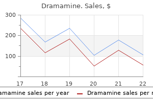
Cheap dramamine 50mg line. Dizziness Lightheadedness & Off Balance - Anxiety Symptoms 101.
Delta Dental Policy None D5988 Surgical splint None D5991 Vesiculobullous disease medicament carrier Not to be used for fluoride gel or bleach symptoms of buy generic dramamine 50mg on-line. Used as a vehicle to deliver prescribed medicaments for sustained contact with the gingiva symptoms 4dp5dt fet generic dramamine 50mg with mastercard, alveolar mucosa treatment nail fungus best dramamine 50mg, and into the periodontal sulcus or pocket Used for procedure that is not adequately described by a code treatment 1860 neurological generic 50 mg dramamine mastercard. General Policy - For benefits purposes, anesthesia is an integral part of the procedures being performed and additional fees are not billable to the patient. General Policy - Benefits are denied, unless covered by the group/individual contract. D6012 Surgical placement of interim Includes removal during later Benefits are denied. An eposteal (subperiosteal) D6040 Surgical placement: eposteal None implant framework of a biocompatible material designed and fabricated to fit on the surface of the bone of the mandible or maxilla with permucosal extensions which provide support and attachment of a prosthesis. Transosteal implants are placed completely through the bone and into the oral cavity from extraoral or intraoral. Modification of a prefabricated D6056 Prefabricated abutment includes modification and abutment may be necessary. A single metal-ceramic crown restoration that is retained, supported and stabilized by an abutment on an implant. A single cast metal crown restoration that is retained, supported and stabilized by an abutment on an implant. A single crown restoration that is retained, supported and stabilized by an implant. A single metal-ceramic crown restoration that is retained, supported and stabilized by an implant. A ceramic retainer for a fixed partial denture that gains retention, support and stability from an abutment on an implant. This is not a per implant code, and is indicated for implant supported fixed prostheses. Fees are not billable to the patient when performed in the same quadrant by the same dentist/dental office as D4341/D4342 or D4240/D4241, D4260/D4261 or D6101/D6102. Fees for retreatment by the same dentist/dental office within 24 months of initial therapy are not billable to the patient. Fees are not billable to the patient when performed within 12 months of restoration (D6058-D6077, D6085, D6094, D6118, D6119, D6194) placement by same dentist/dental office. Fees are not billable to the patient when performed in conjunction with D1110, D4346 or D4910. None D6082 implant supported crown porcelain fused to predominantly base alloys implant supported crown porcelain fused to noble alloys D6083 A single metal-ceramic crown restoration that is retained, supported and stabilized by an implant. A single noble metal-ceramic crown restoration that is retained, supported and stabilized by an implant. A single metal crown restoration that is retained, supported and stabilized by an implant. Used when a period of healing is necessary prior to fabrication and placement of permanent prosthetic. This procedure involves the repair or replacement of any part of the implant supported prosthesis. This procedure applies to the replaceable male or female component of the attachment. Delta Dental Policy None D6086 D6087 D6088 D6085 None None None Benefits are denied. Fees for recementation or rebonding of crowns are not billable to the patient if done within six months of the initial seating date by the same dentist/dental office. Benefit when billed by a dentist/dental office other than the one who seated the crown or performed the previous recementation or rebonding. Fees for recementation or rebonding of fixed partial dentures are not billable to the patient if done within six months of the initial seating date by the same dentist/dental office. Benefit one recementation or rebonding after six months have elapsed since the initial placement. Subsequent requests for recementation or rebonding by the same dentist/dental office are denied. Benefit when billed by a dentist other than the one who seated the crown or performed the previous recementation or rebonding None D6093 Recement or rebond implant/abutment supported fixed partial denture None D6094 Abutment supported crown titanium or titanium alloys D6095 Repair implant abutment, by report A single restoration that is retained supported and stabilized by an abutment on an implant. This procedure involves the repair or replacement of any part of the implant abutment. A metal-ceramic retainer for a fixed partial denture that gains retention, support, and stability from an abutment on an implant. A metal-ceramic retainer for a fixed partial denture that gains retention, support, and stability from an implant. Fees are not billable to the patient when performed in the same surgical site by the same dentist/dental office on the same date of service as D6102. Fees are not billable to the patient when billed in conjunction with D4260 or D4261. Any items in the nomenclature listed separately should be not billable to the patient in conjunction with this procedure. Placement of a barrier membrane or biologic materials to aid in osseous regeneration are reported separately. Bone graft at time of placement Placement of a barrier membrane or biologic materials to aid in osseous regeneration, are reported separately. Benefits billed in conjunction with implants, implant removal, ridge augmentation or preservation, in extraction sites, periradicular surgery, etc. D6110 Implant /abutment supported removable denture for edentulous arch - maxillary Implant /abutment supported removable denture for edentulous arch - mandibular Implant /abutment supported removable denture for partially edentulous arch - maxillary Implant /abutment supported removable denture for partially edentulous arch - mandibular Implant /abutment supported fixed denture for edentulous arch - maxillary Implant /abutment supported fixed denture for edentulous arch - mandibular Placement of a barrier membrane, or biologic materials to aid in osseous regeneration are reported separately. None D6111 None D6112 None D6113 None D6114 None D6115 None When billed in conjunction with implants, implant removal, ridge augmentation or preservation, in extraction sites, periradicular surgery, etc. A metal retainer for a fixed partial denture that gains retention, support, and stability from an implant. Policies in this Handbook that address benefits, limitations and exclusions are policies that have not been tailored to reflect the specific terms of applicable group/individual contracts. General Policy - Fees for fixed partial denture prosthetic procedures include the routine use of temporary prosthetics during the time for normal laboratory fabrication of the completed prosthesis and are not billable to the patient when reported within six months. General Policy - For benefit purposes, anesthesia is an integral part of the procedures being performed and additional fees are not billable to the patient. General Policy - Benefits will be based on the number of pontics necessary for the space, not to exceed the normal complement of teeth. General Policy - A posterior fixed bridge and partial denture are not benefits in the same arch within the frequency limitations. General Policy - Fixed prosthodontics are not a benefit for children under 16 years of age. General Policy - Prosthetics (fixed) are subject to a contractual time limitation for replacement. General Policy - the fees for indirectly fabricated restorations and prosthetic procedures include all models, temporaries, laboratory charges and materials, and other associated procedures. Any fees charged for these procedures by the same dentist/dental office in excess of the approved amounts for the indirectly fabricated restorations or prosthetic procedures are not billable to the patient on same date of service. General Policy - Multi-stage procedures are reported and benefited upon completion. The completion date for immediate dentures is the date that the remaining teeth are removed and the denture is inserted. The completion date for fixed partial dentures and crowns, onlays, and inlays is the cementation date regardless of the type of cement utilized. None D6602 None D6603 None None D6604 None None D6605 None None D6606 None None D6607 None None D6608 D6609 D6610 None None None the benefit is limited to conventional fixed prosthetics. Additional abutments necessary due to special conditions or for splinting are optional and if performed should be done with the agreement of the patient to assume the additional cost. Retainer crown - indirect resin Not to be used as a temporary the benefit is limited to D6721 - crown - resin D6710 based composite or provisional prosthesis. None Fees for a provisional retainer crown by the same dentist/dental office that placed the permanent prostheses are not billable to the patient.
Metanephroi Metanephroi-the primordia of permanent kidneys-begin to develop early in the fifth week and start to function approximately 4 weeks later symptoms gluten intolerance order dramamine 50 mg with mastercard. A mature fetus swallows several hundred milliliters of amniotic fluid every day medications for migraines order dramamine 50 mg visa, which is absorbed by its intestines treatment kidney infection generic dramamine 50mg without a prescription. The fetal waste products are transferred through the placental membrane into the maternal blood for elimination by the maternal kidneys medications derived from plants dramamine 50mg free shipping. As it elongates, the metanephric diverticulum penetrates the metanephrogenic blastema-a mass of mesenchyme (see. The stalk of the metanephric diverticulum becomes the ureter, and the cranial portion of the diverticulum undergoes repetitive branching events, forming the branches which differentiate into the collecting tubules of the metanephros (see. The first four generations of tubules enlarge and become confluent to form the major calices (see. The end of each arched collecting tubule induces clusters of mesenchymal cells in the metanephrogenic blastema to form small metanephric vesicles. The tubules differentiate into proximal and distal convoluted tubules, and the nephron loop (Henle loop), together with the glomerulus and its capsule, constitute a nephron (see. B, Transverse section of the embryo showing the position of the intermediate mesenchyme before lateral folding of the embryo. D, Transverse section of the embryo after the commencement of folding, showing the nephrogenic cords. F, Transverse section of the embryo showing the lateral folds meeting each other ventrally. The mesonephric tubules have been pulled laterally; their normal position is shown in A. Figure 12-3 Dissection of the thorax, abdomen, and pelvis of an embryo at Carnegie stage 22, approximately 54 days. Observe the large suprarenal (adrenal) glands and the elongated mesonephroi (mesonephric kidneys). The phallus will develop into a penis or a clitoris depending on the genetic sex of the embryo. The lobulation usually disappears during infancy as the nephrons increase and grow. At term, nephron formation is complete, with each kidney containing 400,000 to 2,000,000 nephrons. The increase in kidney size after birth results mainly from the elongation of the proximal convoluted tubules as well as an increase of interstitial tissue. Although glomerular filtration begins at approximately the ninth fetal week, functional maturation of the kidneys and increasing rates of filtration occur after birth. C to F, Successive stages in the development of a mesonephric tubule between the 5th and 11th weeks. Note that the mesenchymal cell cluster in the nephrogenic cord develops a lumen, thereby forming a mesonephric vesicle. The vesicle soon becomes an S-shaped mesonephric tubule and extends laterally to join the mesonephric duct. The expanded medial end of the mesonephric tubule is invaginated by blood vessels to form a glomerular capsule. B to E, Successive stages in the development of the metanephric diverticulum (fifth to eighth weeks). Observe the development of the ureter, renal pelvis, calices, and collecting tubules. B and C, Note that the metanephric tubules, the primordia of the nephrons, become continuous with the collecting tubules to form uriniferous tubules. D, Observe that nephrons are derived from the metanephrogenic blastema and that the collecting tubules are derived from the metanephric diverticulum. Branching of the metanephric diverticulum is dependent on induction by the metanephric mesenchyme. The metanephric diverticulum and the metanephrogenic blastema interact and induce each other, a process known as reciprocal induction, to form the permanent kidneys. Molecular studies, especially knockout and transgenic analyses in the mouse, show that this process involves two principal signaling systems that use conserved molecular pathways. Recent research has provided insight into the complex interrelated molecular events regulating the development of the kidneys. Transformation of the metanephric mesenchyme to the epithelial cells of the nephron-mesenchymal-epithelial transition-is regulated by mesenchyme factors including Wnt4. As the abdomen and pelvis grow, the kidneys gradually come to lie in the abdomen and move farther apart (see. In effect, the caudal part of the embryo grows away from the kidneys so that they progressively occupy more cranial levels. Initially the hilum of the kidney, where vessels and nerves enter and leave, faces ventrally; however, as the kidney relocates (ascends), it rotates medially almost 90 degrees. Eventually the kidneys become retroperitoneal (external to the peritoneum) on the posterior abdominal wall. Changes in Blood Supply of the Kidneys page 249 page 250 Figure 12-8 the kidneys and suprarenal glands of a 28-week fetus (Г-2). The external evidence of the lobes usually disappears by the end of the first postnatal year. During the changes in kidney position, they receive their blood supply from vessels that are close to them. When they are located at a higher level, they receive new branches from the aorta (see. Normally the caudal branches of the renal vessels undergo involution and disappear. The position of the kidneys becomes fixed once they come into contact with the suprarenal glands in the ninth week. Accessory (supernumerary) renal arteries usually arise from the aorta superior or inferior to the main renal artery and follow the main renal artery to the hilum of the kidney. Accessory renal arteries may also enter the kidneys directly, usually into the superior or inferior poles. An accessory artery to the inferior pole (polar renal artery) may cross anterior to the ureter and obstruct it, causing hydronephrosis-distention of the renal pelvis and calices with urine (see. If the artery enters the inferior pole of the right kidney, it usually crosses anterior to the inferior vena cava and ureter. It is important to be aware that accessory renal arteries are end arteries; consequently, if an accessory artery is damaged or ligated, the part of the kidney supplied by it will become ischemic. C and D, Note that as the kidneys relocate, they are supplied by arteries at successively higher levels and that the hilum of the kidney is eventually directed anteromedially. The polar renal artery, illustrated in B, has obstructed the ureter and produced an enlarged renal pelvis. B, Transverse scan at a slightly higher level showing the left suprarenal gland (between cursors) within the left renal fossa. The small sketch to the lower right of each drawing illustrates the probable embryologic basis of the anomaly. C, Right side, malrotation of the kidney; left side, bifid ureter and supernumerary kidney. E, Discoid kidney resulting from fusion of the kidneys while they were in the pelvis. F, Supernumerary left kidney resulting from the development of two metanephric diverticula. This pelvic kidney resulted from its failure to ascend during the sixth to ninth weeks. B, Contrast-enhanced computed tomography scan of the abdomen of an infant with a horseshoe kidney. Note the isthmus (vascular) of renal tissue (I) connecting the right and left kidneys just anterior to the aorta (arrow) and inferior vena cava. Many fetal urinary tract abnormalities can be detected before birth by ultrasonography. Integration link: Antenatal diagnosis of urinary tract anomalies Renal Agenesis Unilateral renal agenesis occurs approximately once in every 1000 newborn infants. Males are affected more often than females, and the left kidney is usually the one that is absent.
References