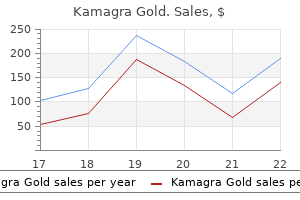
X


|
STUDENT DIGITAL NEWSLETTER ALAGAPPA INSTITUTIONS |

|
Paul Berkowitz, MD
Relationship of retinal vascular tortuosity with the neuroretinal rim: the Singapore Malay Eye Study erectile dysfunction blog buy cheap kamagra gold 100mg on line. Refractive error drinking causes erectile dysfunction cheap kamagra gold 100mg line, axial dimensions erectile dysfunction drugs stendra buy cheap kamagra gold 100 mg line, and primary open-angle glaucoma: the Singapore Malay Eye Study best male erectile dysfunction pills buy 100 mg kamagra gold. Relationship of Central Corneal Thickness with Optic Disc Parameters: the Singapore Malay Eye Study. Distribution of ocular perfusion pressure and its relationship with open-angle glaucoma: the Singapore Malay Eye Study. Ocular and systemic factors related to intraocular pressure in Japanese adults: the Tajimi study. Population prevalence of tilted and torted optic discs among an adult Chinese population in Singapore: the Tanjong Pagar Study. Risk factors for primary open-angle glaucoma and pseudoexfoliative glaucoma in the Thessaloniki Eye Study. Association of blood pressure status with the optic disk structure in non-glaucoma subjects: the Thessaloniki Eye Study. Factors associated with undiagnosed openangle glaucoma: the Thessaloniki Eye Study. Increased likelihood of glaucoma at the same screening intraocular pressure in subjects with pseudoexfoliation: the Thessaloniki Eye Study. The tube versus trabeculectomy study: design and baseline characteristics of study patients. Treatment outcomes in the Tube Versus Trabeculectomy Study after one year of follow-up. Surgical complications in the Tube Versus Trabeculectomy Study during the first year of follow-up. Electrophysiologic Testing in Disorders of the Retina, Optic Nerve, and Visual Pathway (American Academy of Ophthalmology Monograph Series) by Gerald Allen Fishman, David G. Clinical Ophthalmic Pathology: Principles of Diseases of the Eye and Associated Structures. Stereoatlas of Ophthalmic Pathology: Anatomy and Pathology of the Peripheral Fundus (Fundus extremus). American Academy of Ophthalmology: Basic and Clinical Science Course 2011-2012 Section 4: Ophthalmic Pathology and Intraocular Tumors. Biswas J, Krishnakumar S, Ahuja S: Manual of ocular pathology, Jaypee Brothers, India, 2010. Ophthalmic plastic surgery: decision making and techniques New York: McGraw-Hill, Medical Publishing Div; 2002. Ophthalmic and facial plastic surgery: a compendium of reconstructive and aesthetic techniques. Surgery of the eyelid, orbit, and lacrimal system (3 vols) (Ophthalmology monographs 8). Symposium on Plastic Surgery in the Orbital Region, Dallas, 1974 (Proceedings of the Symposium of the Educational Foundation of the American Society of Plastic and Reconstructive Surgeons; v. Fundamental Techniques of Plastic Surgery and their Surgical Applications, Churchill Livingstone. Jeong S, et al: the Asian Upper Eyelid: An Anatomical Study with Comparison to the Caucasian Eyelid. Tessier P: Anatomical classification of facial, cranio-facial, and latero-facial clefts. American Academy of Ophthalmology: Functional indications for upper and lower eyelid blepharoplasty. Faigen S: Advanced Rejuvenative Upper Blepharoplasty: Enhancing Aesthetics of the Upper Periorbita. Esmaeli B: Sentinel lymph node mapping for patients with cutaneous and conjunctival malignant melanoma. Esmaeli B, Wang B, Deavers M, Gillenwater A, et al: Prognostic factors for survival in malignant melanoma of the eyelid skin. Shorr N, Seiff S: the four stages of surgical rehabilitation of the patient with dysthyroid ophthalmopathy. Shorr N, Neuhaus R, Baylis H: Ocular motility problems after orbital decompression for dysthyroid ophthalmopathy. Kazim M: Radiotherapy for Graves ophthalmopathy: the Columbia University experience. Rituximab treatment of patients with severe, corticosteroid-resistant thyroid-associated ophthalmopathy: Ophthalmology. Kohn R, Hepler R: Management of limited rhino-orbital mucormycosis without exenteration. Liu D: A simplified technique of orbital decompression for severe retrobulbar hemorrhage. Wharam M et al: Localized orbital rhabdomyosarcoma: an interim report of the intergroup rhabdomyosarcoma study committee. Beard C: Dermolipoma surgery, or, "An ounce of prevention is worth a pound of cure. Smith B, Petrelli R: Dermis -fat graft as a moveable imp lant within the muscle cone. Hartikainen J, Crenman R, Puukka P, Seppa H: Prospective Randomized Comparison of External Dacryocystorhinostomy and Endonasal Laser Dacryocystorhinostomy. Nonlaser endoscopic endonasal dacryocystorhinostomy with adjunctive mitomycin C in nasolacrimal duct obstruction in adults. De Groot V, De Wilde F, Smet L, Tassignon M-J: Frontalis Suspension Combined with Blepharoplasty as an Effective Treatment for Blepharospasm Associated with Apraxia of Eyelid Opening. Zaturansky B, Hyams S: Perforation of the globe during the injection of local anesthesia. Task Force on Sedation and Analgesia in Ambulatory Settings: Sedation and Analgesia in Ambulatory Settings. Fagien S: Botox for the treatment of dynamic and hyperkinetic facial lines and furrows: adjunctive use in facial surgery. Patipa M: the Evaluation and Management of Lower Eyelid Retraction Following Cosmetic Surgery. The Strabismus Minute Basic Examination Techniques for Children and Adults with Strabismus 1. Artifacts introduced by spectacle lenses in the measurement of strabismic deviations. In: Symposium on strabismus: Transactions of the New Orleans Academy of Ophthalmology. The deterioration of accommodative esotropia: Frequency, characteristics, and predictive factors. Visual acuity results following treatment of persistent hyperplastic primary vitreous. Randomized trial to evaluate combined patching and atropine for residual amblyopia. Treatment of severe amblyopia with weekend atropine: results from 2 randomized clinical trials. Eye muscle surgery for Infantile Nystagmus syndrome in the first two years of life. Long-term results of adjustable suture surgery for strabismus secondary to thyroid ophthalmopathy. Simultaneous correction of blepharoptosis and exotropia in aberrant regeneration of the oculomotor nerve by strabismus surgery. In: Pediatric Ophthalmology and Strabismus: Transactions of the New Orleans Academy of Ophthalmology. Classification and surgical treatment of superior oblique palsies: I Unilateral superior oblique palsies. In: Pediatric Ophthalmoloy and Strabismus: Transactions of the New Orleans Academy of Ophthalmology. Vertical Offsets of horizontal recti muscles in the management of A and V pattern strabismus.
G encore vacuum pump erectile dysfunction cheap kamagra gold 100mg, the problem with this splint is that it was intended to be used for only a few days impotence treatment devices kamagra gold 100mg without prescription, but the patient wore it and walked on it for 3 wk erectile dysfunction zocor 100 mg kamagra gold free shipping. The posterior splint is used primarily to immobilize severe ankle sprains erectile dysfunction remedy generic kamagra gold 100mg visa, fractures of the distal fibula and tibia, and reduced ankle dislocations. It can also be used for fractures of the tarsal and metatarsal bones or for other foot conditions that require immobilization. In particularly severe or unstable injuries, an additional anterior splint may be used to provide extra immobilization resembling that of a formal cast (. For severe lateral or bilateral ligamentous injuries, a U-splint or stirrup splint (see later) may be added to the posterior splint for increased immobilization. With minor soft tissue injuries, patients may have partial weight-bearing on ankle splints after 24 hours. If the patient will be bearing weight, a cast shoe over the splint makes it easier to walk. In addition, a cast shoe increases the longevity of the splint because walking on an unprotected splint quickly destroys the device. Generally, walking on the splint is prohibited if immobilization for more than 2 or 3 days is desired. In particularly severe or unstable injuries, an anterior splint may be added to a posterior ankle splint to provide extra immobilization resembling that of a formal cast. The anterior splint is never used by itself, but it can augment a posterior splint, creating a bivalve effect. It should extend from the plantar surface of the great toe or metatarsal heads along the posterior surface of the foreleg to the level of the fibular head. If it hurts to move the toes, they should be incorporated into the splint (after padding is placed between the digits). It is a common mistake to apply a posterior splint that does not extend far enough to support the ball of the foot. Fifteen to 20 layers should be used if partial weight-bearing is allowed because this splint frequently breaks or cracks when walked on. With the knee and ankle in the proper position, stockinette may be applied and the foot and leg padded with Webril, as described earlier. Again, Webril or gauze is placed between the toes if they are to be included in the splint. The wet plaster is then laid over the plantar surface of the foot and secured in place by folding back the ends of the stockinette and wrapping with one or two 4-inch-wide elastic bandages. The wet plaster is carefully molded around the malleoli and instep to ensure maximum comfort and immobilization. The toes should be left partially exposed for later examination of color and capillary refill. Anterior-Posterior Splint Indications the anterior splint is never used by itself, but it can augment a posterior splint, creating a bivalve effect (see. Construction A piece of plaster should be cut several centimeters shorter than the one used for the posterior splint, but because this splint does not bear weight, only 8 to 10 layers are required. Application the patient should be positioned and padded as for the posterior splint. After the wet posterior splint has been applied, the anterior splint is placed over the anterior aspect of the ankle and foreleg parallel to the posterior splint. The two are then held in place with elastic bandages as described earlier for the posterior splint alone. An assistant is needed to apply the anterior-posterior splint because it is extremely difficult to hold both splints in place while wrapping the elastic bandages. It functions like the posterior splint, and either of the two provides satisfactory ankle immobilization. In one study that compared these splints in normal volunteers, the U-splint allowed less plantar flexion and broke less often with plantar flexion than the posterior splint. The splint passes under the plantar surface of the foot, extending up the medial and lateral sides of the foreleg to just below the level of the fibular head. For immobilization of the knee, the sides of the splint may be extended proximally to the groin, creating a long leg splint. The splint passes under the plantar surface of the foot from the calcaneus to the metatarsal heads and extends up the medial and lateral sides of the foreleg to just below the level of the fibular head. Application the patient is positioned, and the extremity is padded as described for the posterior splint. The wet plaster is laid across the plantar surface of the foot between the calcaneus and the metatarsal heads with the sides extending up the lateral and medial aspects of the foreleg. The elastic bandage should be wrapped around the extremity starting at the metatarsal heads and continuing around the ankle using a figure-of-eight configuration. Once the ankle has been wrapped, another 4- or 6-inch elastic bandage can be used to secure the remainder of the splint in place. The walking boot provides a similar degree of immobilization as a U-splint, but is easier to remove for bathing and dressing, and the Velcro straps allow adjustment for edema. B, the Unna boot or an Ace wrap provide effective immobilization of an ankle soft tissue injury. For similar short-term immobilization without plaster, a modified Jones dressing can be used. Copious Webril is wrapped around the ankle and foot and covered with an elastic bandage. When cleared by the follow-up physician, the walking boot allows easy transition to full weight-bearing. Studies have shown that rapid mobilization after ankle injuries improves functional outcome and reduces disability time. Once the appropriate size has been determined, the patient places her or his bare foot and ankle into the boot, which is secured using Velcro straps. Semirigid Orthosis Indications In patients with sprains of the lateral ankle associated with a stable joint, the use of a functional brace with early mobilization is frequently more comfortable, and results in an earlier return to normal function, than complete 22 immobilization in a plaster splint or cast. However, it should be pointed out that there is no documented difference in long-term outcome between the two methods of treatment. Application Most functional ankle braces resemble a U-splint with air bladders (Aircast, Inc. Hard Shoe (Cast or Reese Shoe) Indications A hard shoe can help reduce the pain associated with ambulation in patients with fractures or soft tissue injuries to the foot. This device can also be used over a splint or cast to allow partial weight-bearing. Application If the cast shoe is going to be used by a patient with a fractured toe, the injured digit should first be buddy taped to the adjacent toe. Ankle Wraps and Bandages There are no data supporting the routine use of ankle wraps for simple sprains, but some pain relief may be afforded by a proper wrap. It should not be tight enough to impair venous drainage, a common problem when patients apply their own wraps. To apply a Jones dressing, generously wrap the foot and ankle with large amounts of Webril (about five or seven layers) and cover it with an elastic bandage. Soft Cast Indications A soft cast is basically a modified Jones compression dressing. It is useful for minor ligamentous and soft tissue injuries of the foot and ankle that do not require prolonged or complete immobilization. A soft cast can help reduce the pain and swelling often associated with mild ankle sprains and gives support for early weight-bearing (see. To begin, the patient is placed in a supine position with the foot and ankle extending off the end of the stretcher. Alternatively, the leg can be elevated by an assistant or by placing pillows under the knee and foreleg. The ankle and foot are then wrapped with five or seven layers of Webril, starting at the metatarsal heads and continuing around the ankle in a figure-of-eight configuration.

The head of the tonometer then automatically presses against the cornea erectile dysfunction treatment jaipur generic 100 mg kamagra gold free shipping, measures intraocular pressure erectile dysfunction age 36 purchase kamagra gold 100mg fast delivery, and retracts erectile dysfunction pills in south africa buy kamagra gold 100mg line. A patient tonometer may be prescribed in applicable cases (such as increased risk of acute glaucoma) treatment of erectile dysfunction using platelet-rich plasma buy generic kamagra gold 100 mg on-line. However, using the device requires a certain degree of skill on the part of the patient. Patients who have problems applying eyedrops are best advised not to attempt to use a patient tonometer. Younger and well motivated patients are the best candidates for tonometric self-examination. In the presence of persistently elevated intraocular pressure, the optic cup becomes enlarged and can be evaluated by ophthalmoscopy. Stereoscopic examination of the optic disk through a slit-lamp biomicroscope fitted with a contact lens provides a three-dimensional image. Large normal optic cups are nearly always round and differ from the vertical elongation of the optic cup seen in eyes with glaucoma. Documenting the optic disk: Recording findings in sketches is suitable for routine documentation and follow-up examination of the optic disk. Optic disk measurement and tomography can provide precise measurements of the optic nerve. The area of the optic disk, optic cup, and neuroretinal rim (vital optic disk tissue) can be measured by planimetry on two-dimensional photographs of the optic nerve. Modern laser scanning ophthalmoscopes permit three-dimensional documentation of the optic nerve. Glaucomatous changes in the optic nerve: Glaucoma produces typical changes in the shape of the optic cup. Progressive destruction of nerve fibers, fibrous and vascular tissue, and glial tissue will be observable. This tissue atrophy leads to an increase in the size of the optic cup and to pale discoloration of the optic disk. Progressive glaucomatous changes in the optic disk are closely associated with increasing visual field defects. We know that glaucomatous visual field defects initially manifest themselves in the superior paracentral nasal visual field or, less frequently, in the inferior field, as relative scotomas that later progress to absolute scotomas. The computer then calculates crucial data for the optic disk and presents a stereometric analysis (d). The blood vessels abruptly plunge into the deep cup, indicated by their typical bayonetshaped kinks in the image (arrow). Computer-controlled semiautomatic grid perimetry devices such as the Octopus or Humphrey field analyzer are used to examine the central 30 degree field of vision (modern campimetry;. Reproducible visual field findings are important in follow-up to exclude any enlargement of the defects. Peripheral optic cup in a temporal and inferior location (with damage to the optic nerve fibers in this area). Advanced generalized thinning of the neuroretinal rim with an increasingly visible lamina cribrosa and nasal displacement of the blood vessels. Total glaucomatous atrophy of the optic nerve: Complete atrophy of the neuroretinal rim, kettleshaped optic cup, bayonet kinks in the blood vessels on the margin of the optic disk, some of which disappear. The optic disk is surrounded by a ring of chorioretinal atrophy (glaucomatous halo) due to pressure atrophy of the choroid and lysis of the retinal pigmented epithelium. The arc-shaped scotoma has expanded into a ring-shaped scotoma surrounding the focal point. As the focal point degenerates, the center of vision disappears and only a peripheral residual field of vision remains. The standardized examination conditions in automatic perimetry not only permit early detection of glaucoma; the reproducible results also aid in the prompt diagnosis of worsening findings. In addition to the early progressive optic nerve and visual field defects, arcshaped defects also occur in the nerve fiber layer. The angle of the anterior chamber characteristically remains open throughout the clinical course of the disorder. Epidemiology: Primary open angle glaucoma is by far the most common form of glaucoma and accounts for over 90% of adult glaucomas. The incidence of the disorder significantly increases beyond the age of 40, reaching a peak between the ages of 60 and 70. Patients with a positive family history are at greater risk of developing the disorder. Etiology (See also physiology and pathophysiology of aqueous humor circulation): the cause of primary open angle glaucoma is not known, although it is known that drainage of the aqueous humor is impeded. The primary lesion occurs in the neuroretinal tissue of the optic nerve as compression neuropathy of the optic nerve. Symptoms: the majority of patients with primary open angle glaucoma do not experience any subjective symptoms for years. However, a small number of patients experience occasional unspecific symptoms such as headache, a burning sensation in the eyes, or blurred or decreased vision that the patient may attribute to lack of eyeglasses or insufficient correction. The patient may also perceive rings of color around light sources at night, which has traditionally been regarded as a symptom of angle closure glaucoma. Primary open angle glaucoma can be far advanced before the patient notices an extensive visual field defect in one or both eyes. It is crucial to diagnose the disorder as early as possible because the prognosis for glaucoma detected in its early stages is far better than for advanced glaucoma. Where increased intraocular pressure remains undiagnosed or untreated for years, glaucomatous optic nerve damage and the associated visual field defect will increase to the point of blindness. Elevated intraocular pressure in a routine ophthalmic examination is an alarming sign. The angle of the anterior chamber is open and appears as normal as the angle in patients without glaucoma. Examination of the optic nerve reveals whether glaucomatous cupping has already occurred and how far advanced the glaucoma is. Where the optic disk and visual field are normal, ophthalmoscopic examination of the posterior pole under green light may reveal fascicular nerve fiber defects as early abnormal findings. Noise field perimetry is suitable as a screening test as it makes the patient aware of scotomas and makes it possible to detect and describe them. The patient is shown a flickering monitor displaying what resembles image noise on a television set. In advanced glaucoma, kinetic hand perimetry with the Goldmann perimeter device is a useful preliminary examination to evaluate the remaining field of vision. Differential diagnosis: Two disorders are important in this context: Ocular hypertension. Patients with ocular hypertension have significantly increased intraocular pressure over a period of years without signs of glaucomatous optic nerve damage or visual field defects. Some patients in this group will continue to have elevated intraocular pressure but will not develop glaucomatous lesions; the others will develop primary open angle glaucoma. The probability that a patient will develop definitive glaucoma increases the higher the intraocular pressure, the younger the patient, and the more compelling the evidence of a history of glaucoma in the family. Patients with low-tension glaucoma exhibit typical progressive glaucomatous changes in the optic disk and visual field without elevated intraocular pressure. These patients are very difficult to treat because management cannot focus on the control of intraocular pressure. Often these patients will have a history of hemodynamic crises such as gastrointestinal or uterine bleeding with significant loss of blood, low blood pressure, and peripheral vascular spasms (cold hands and feet). Patients with glaucoma may also experience further worsening of the visual field due to a drop in blood pressure. Caution should be exercised when using cardiovascular and anti-hypertension medications in patients with glaucoma. O Glaucomatous changes in the optic cup: Medical treatment should be initiated where there are signs of glaucomatous changes in the optic cup or where there is a difference of more than 20% between the optic cups of the two eyes. O Increasing glaucomatous changes in the optic cup or increasing visual field defects: Regardless of the pressure measured, these changes show that the current pressure level is too high for the optic nerve and that additional medical therapy is indicated.

As recently as the turn of the 20th century erectile dysfunction shots discount kamagra gold 100 mg fast delivery, however icd 9 code erectile dysfunction 2011 discount 100 mg kamagra gold visa, malaria and typhoid were major health problems erectile dysfunction doctor washington dc cheap 100mg kamagra gold with visa. Flies erectile dysfunction pills herbal discount 100 mg kamagra gold otc, the order Diptera, are one of the largest and most dynamic orders of insects. Most flies are small and soft-bodied with two large eyes on the front of the head. Flies can be divided into two groups, depending on the appearance of the larvae and adults. Their antennae are short or not visible; some are relatively large but usually not long-legged. Their harborage varies-they live in water, filth, soil, carcasses, plant tissues, or animal tissues. To enter a house, they have flown inside through an open door or window, or they have moved from a dead animal in a wall. Their thorax and abdomen are shiny black, metallic green or bronze, or they have a metallic blue abdomen with a dull thorax. They have a dull gray thorax with dark stripes and a dark, dull abdomen with yellow sides. Blowflies: greenbottle fly, Phaenicia sericata (left), and bluebottle fly, Calliphora vicina (right). In favorable weather, housefly larvae mature in 6 to 10 days and blowflies in 3 to 9 days. Flesh flies (the family Sarcophagidae) live on meat scraps, dead animals, and dog excrement. They are more than 1/4 inch long, have a dull gray thorax with three distinct dark stripes, and a gray checkerboard abdomen. Section 3: Chapter 11 100 When any of these flies become problems inside, their breeding site and their larvae will usually be close by. Garbage cans and dumpsters are often the problem source; even soil where garbage has decomposed will support infestations. Blowflies are scavengers and live in manure, carrion, dead birds, and dead rodents in wall voids and chimneys. Look for door props and hooks, as well as gaps where broom handles are stuck over hinges to hold the door open or for doors that do not fit tightly. Non-chemical controls include: s Electric flytraps will control only a low level of adult flies. Do not put them in competition with other lights, such as those from vending machines, etc. Follow-up Regularly check sanitation and exclusion methods to see that they are being maintained. Observe client and worker habits that run counter to the pest management program (sanitation, habitat alteration, and so forth). In nature, overwintering locations are under bark, in hollow parts of trees, or under the bark of logs. If they begin investigating structure walls in their search for winter harborage, their upward movement often brings them to openings under siding, ventilators, and weep holes in masonry, cracks around windows, wire penetrations, wall voids, and openings around the roof. They often make their way down through closets and chimney cracks into living spaces of the house. This same behavior takes place in office buildings, hospitals, and other structures. Make the following recommendations to clients: s Remove breeding materials such as garbage and manure. Use exclusion techniques to prevent flies from entering, such as: s Caulk and tighten around all openings, such as screens, doors, windows, ventilators, and eaves. Pesticide Application s Fly strips can be placed in low-access rooms, such Pesticide Application s Use liquid pressurized sprays or dusts where flies as attics and storerooms. They are about 1/ inch long 8 and somewhat similar looking, but their biology and management are very different. Treatments of these fly infestations are a good example of the site-specific nature of successful pest management. Control and Management of Fruit Flies Inspection When certain the infesting insect is a fruit fly, look for fermenting materials. The head and thorax are yellowish to brown, and the abdomen is light brown to dark with yellow bands. It consists of a thickened vein bordering the front margin of the wing from the attachment at the thorax to the wing tip. In a common fruit fly infestation, flies are attracted to the sweet odor of fermentation in ripe fruit, such as bananas; they lay their eggs in the cracks of the peel. Newly emerged adults are attracted to lights, but egglaying females will not leave fermenting materials. Fruits, vegetables, beer, fermenting water from refrigerators, humidifiers, sink drains, sour mops and rags, and fermenting pet food are examples of fermenting material. Infestations are common in orchards, breweries, restaurants, canneries, hospitals, and homes. They are dark brown and look humpbacked-because the small head is located low on the front bulge of the thorax. Wing venation consists of several short, thickened veins on the fore margin of the wing near the attachment to the thorax. These veins do not extend to the wing tip, and other veins are weak or nearly invisible. Adults are able to emerge from the underground infestation site upward through several feet of soil. If broken Section 3: Chapter 11 102 General Pest Management sewer lines are under buildings, phorids can come up through cracks in concrete floors or around floor drains. When water and sewage wash out cavities in the soil around the pipe, immense numbers of flies are produced. When sewage treatment plant filter beds malfunction or become "out of balance," the moth flies can become problems in nearby neighborhoods. Control and Management of Phorid Flies Inspection Carefully identify the infesting fly as a phorid. Ask clients if there have been sewer problems, buried garbage, or decaying vegetable or animal matter close by. Their larvae infest moist soil and feed on fungi associated with decaying vegetation. They also build up in pigeon droppings on outside ledges, then enter dwellings through nearby windows. Their dark color comes from tiny hairs that cover the wings, which are held in roof-like fashion over the body. When drain traps of sinks, commodes, and floor drains dry out, large numbers can enter dwellings from the sewer. In sewage treatment plants, drain flies feed on the gelatinous material that collects on stones in trickling filter beds. Over time, however, cast skins from these filter General Pest Management 103 Figure 11. Section 3: Chapter 11 Adult midges are often a food source for spiders on buildings and monuments (see Web-weaving Spiders, Chapter 13). These insects are responsible for millions of deaths each year because of their disease-vectoring ability, particularly in less-developed countries. In urban areas, flies contaminate food and people in restaurants, hospitals, and homes. Briefly describe the two major divisions of the order Diptera characterized by form and give an example for each. Cluster flies (along with houseflies, face flies, some blowflies, and flesh flies) are referred to as "attic flies" because they often overwinter as adults in unused attics. List at least three pest management procedures for controlling attic/cluster flies. List at least three pest management procedures for fruit fly or Drosphila infestations.
Purchase kamagra gold 100 mg on line. Wu is on the phone.
References